Free Website Builder. Unlimited Storage. Unlimited Websites
Create New Website

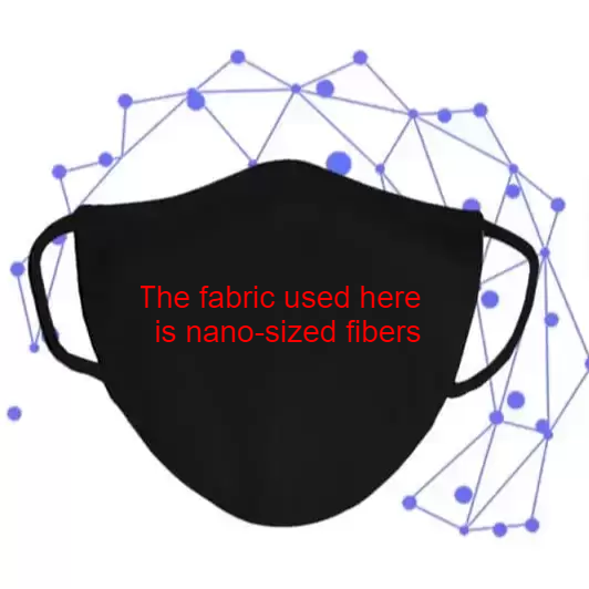
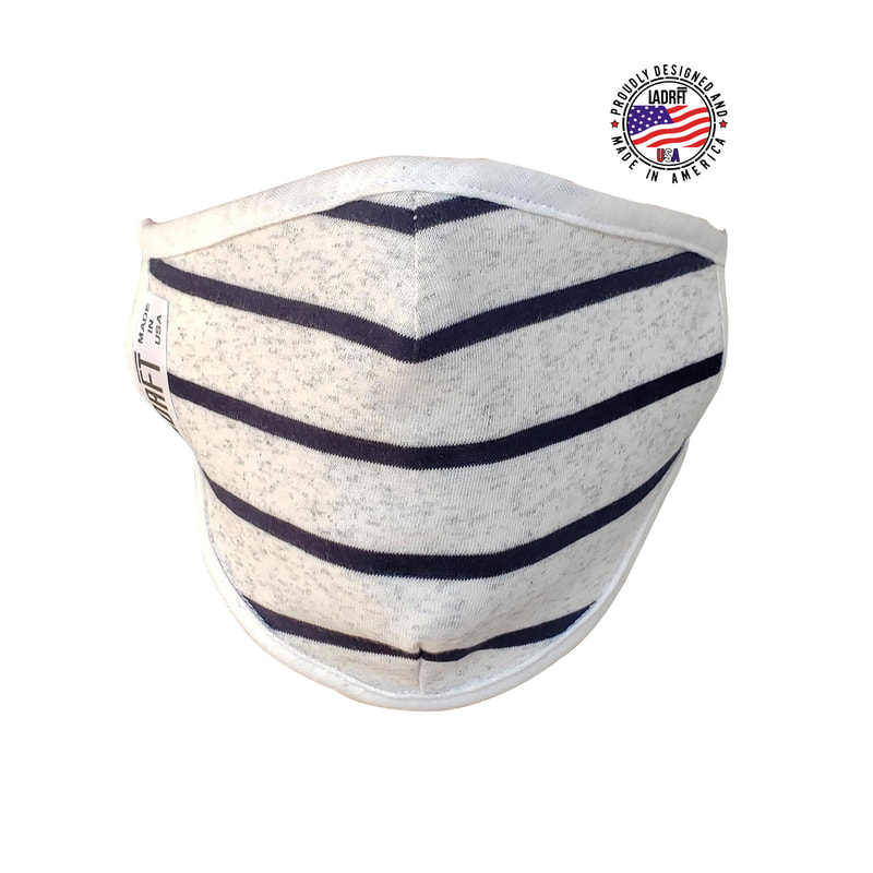
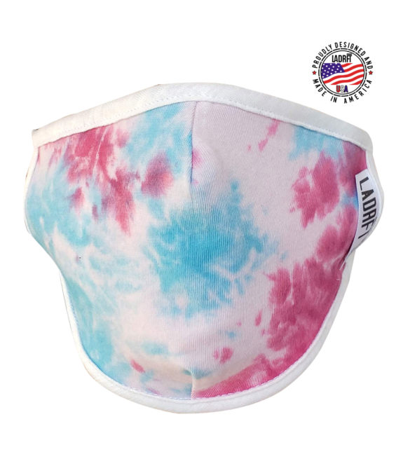
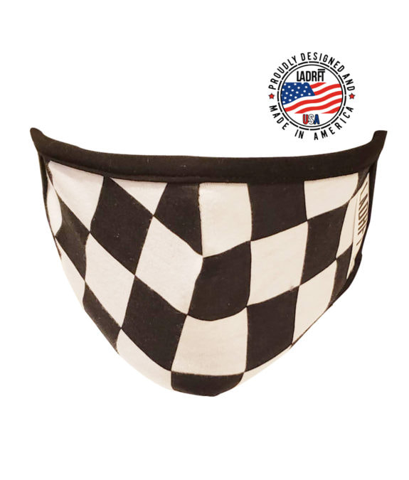
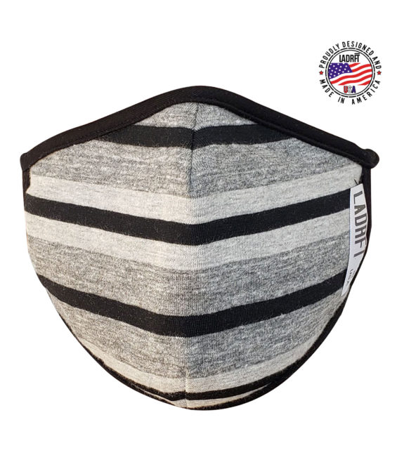
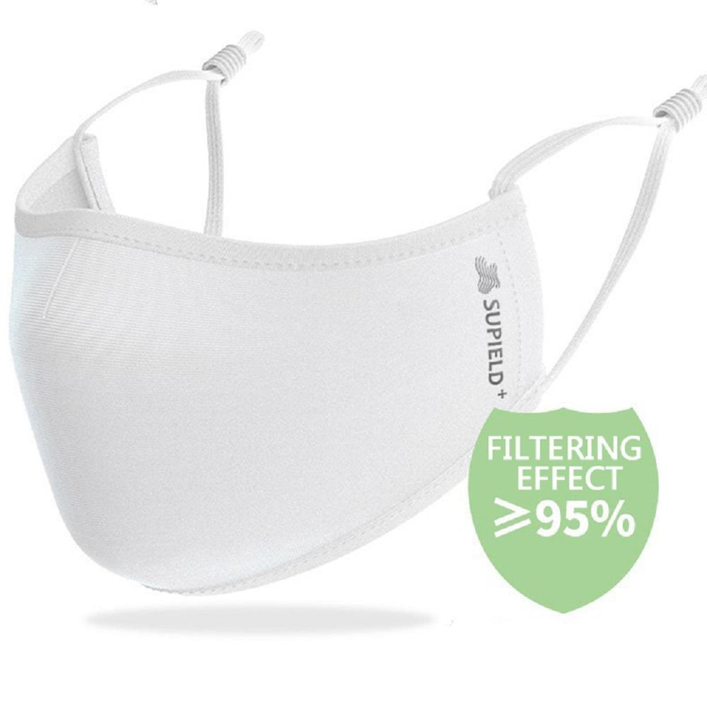
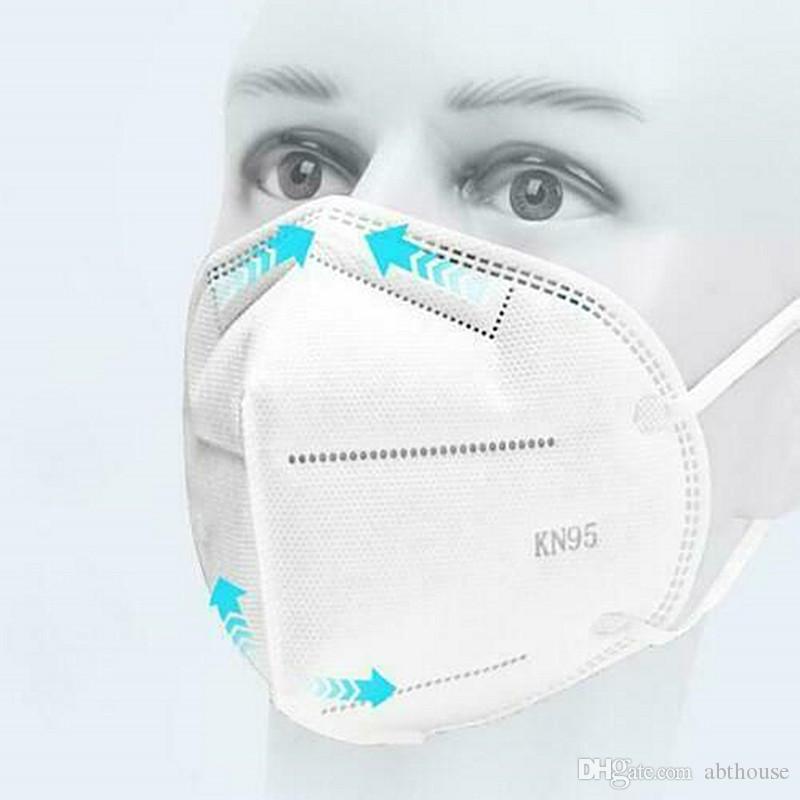
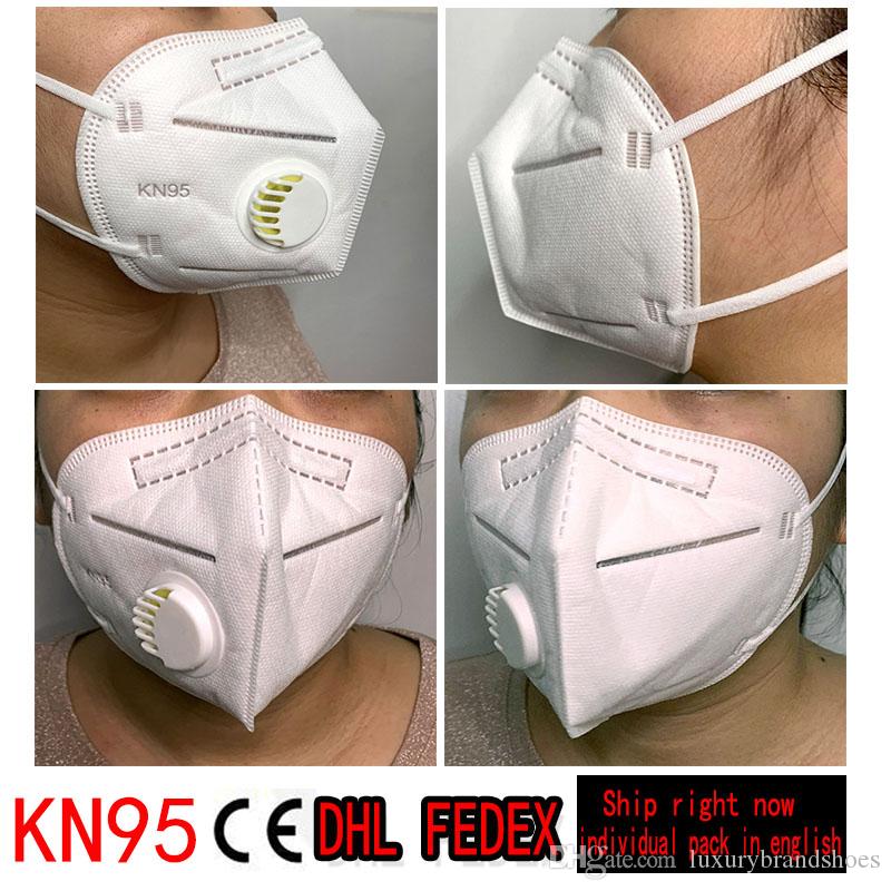
What Makes CREATIVE NANO FABRIC Facemasks to be Above The Competition
COVID-19 Related Information
Comments from use of CREATIVE COVID-19 Facemask


How to Wear Cloth Face Coverings
Cloth face coverings should--

Hypochlorous acid
From Wikipedia, the free encyclopedia
Hypochlorous acid (HOCl) is a weak acid that forms when chlorine dissolves in water, and itself partially dissociates, forming hypochlorite, ClO−. HClO and ClO− are oxidizers, and the primary disinfection agents of chlorine solutions.[2] HClO cannot be isolated from these solutions due to rapid equilibration with its precursor. Sodium hypochlorite (NaClO) and calcium hypochlorite (Ca(ClO)2), are bleaches, deodorants, and disinfectants.
History
Hypochlorous acid was discovered in 1834 by the French chemist Antoine Jérôme Balard (1802–1876) by adding, to a flask of chlorine gas, a dilute suspension of mercury(II) oxide in water.[3] He also named the acid and its compounds.[4]
Uses
Formation, stability and reactions
Addition of chlorine to water gives both hydrochloric acid (HCl) and hypochlorous acid (HOCl):[13]
Cl2 + H2O ⇌ HClO + HCl
Cl2 + 4 OH− ⇌ 2 ClO− + 2 H2O + 2 e−
Cl2 + 2 e− ⇌ 2 Cl−
When acids are added to aqueous salts of hypochlorous acid (such as sodium hypochlorite in commercial bleach solution), the resultant reaction is driven to the left, and chlorine gas is formed. Thus, the formation of stable hypochlorite bleaches is facilitated by dissolving chlorine gas into basic water solutions, such as sodium hydroxide.
The acid can also be prepared by dissolving dichlorine monoxide in water; under standard aqueous conditions, anhydrous hypochlorous acid is currently impossible to prepare due to the readily reversible equilibrium between it and its anhydride:[14]
2 HOCl ⇌ Cl2O + H2O K (at 0 °C) = 3.55×10⁻³ dm3 mol−1
The presence of light or transition metal oxides of copper, nickel, or cobalt accelerates the exothermic decomposition into hydrochloric acid and oxygen:[14]
2 Cl2 + 2 H2O → 4 HCl + O2Chemical reactions
In aqueous solution, hypochlorous acid partially dissociates into the anion hypochlorite ClO−:
HOCl ⇌ ClO− + H+
Salts of hypochlorous acid are called hypochlorites. One of the best-known hypochlorites is NaClO, the active ingredient in bleach.
HOCl is a stronger oxidant than chlorine under standard conditions.
2 HClO(aq) + 2 H+ + 2 e− ⇌ Cl2(g) + 2 H2O E = +1.63 V
HClO reacts with HCl to form chlorine gas:
HOCl + HCl → H2O + Cl2HOCl reacts with ammonia to form monochloramine:
NH3 + HOCl → NH2Cl + H2OHOCl can also react with organic amines, forming N-chloroamines.
Reactivity of HClO with biomolecules
This article relies too much on references to primary sources. Please improve this by adding
secondary or tertiary sources. (January 2020) (Learn how and when to remove this template message)
Hypochlorous acid reacts with a wide variety of biomolecules, including DNA, RNA,[8][15][16][17] fatty acid groups, cholesterol[18][19][20][21][22][23][24][25] and proteins.[21][26][27][28][29][30][31]
Reaction with protein sulfhydryl groups
Knox et al.[29] first noted that HClO is a sulfhydryl inhibitor that, in sufficient quantity, could completely inactivate proteins containing sulfhydryl groups. This is because HClO oxidises sulfhydryl groups, leading to the formation of disulfide bonds[32] that can result in crosslinking of proteins. The HClO mechanism of sulfhydryl oxidation is similar to that of monochloramine, and may only be bacteriostatic, because once the residual chlorine is dissipated, some sulfhydryl function can be restored.[28] One sulfhydryl-containing amino acid can scavenge up to four molecules of HOCl.[31] Consistent with this, it has been proposed that sulfhydryl groups of sulfur-containing amino acids can be oxidized a total of three times by three HClO molecules, with the fourth reacting with the α-amino group. The first reaction yields sulfenic acid (R–SOH) then sulfinic acid (R–SO2H) and finally R–SO3H. Sulfenic acids form disulfides with another protein sulfhydryl group, causing cross-linking and aggregation of proteins. Sulfinic acid and R–SO3H derivatives are produced only at high molar excesses of HClO, and disulfides are formed primarily at bacteriocidal levels.[17] Disulfide bonds can also be oxidized by HClO to sulfinic acid.[32] Because the oxidation of sulfhydryls and disulfides evolves hydrochloric acid,[17] this process results in the depletion HClO.
Reaction with protein amino groups
Hypochlorous acid reacts readily with amino acids that have amino group side-chains, with the chlorine from HClO displacing a hydrogen, resulting in an organic chloramine.[33] Chlorinated amino acids rapidly decompose, but protein chloramines are longer-lived and retain some oxidative capacity.[7][31] Thomas et al.[7] concluded from their results that most organic chloramines decayed by internal rearrangement and that fewer available NH2 groups promoted attack on the peptide bond, resulting in cleavage of the protein. McKenna and Davies[34] found that 10 mM or greater HClO is necessary to fragment proteins in vivo. Consistent with these results, it was later proposed that the chloramine undergoes a molecular rearrangement, releasing HCl and ammonia to form an aldehyde.[35] The aldehyde group can further react with another amino group to form a Schiff base, causing cross-linking and aggregation of proteins.[21]
Reaction with DNA and nucleotides
Hypochlorous acid reacts slowly with DNA and RNA as well as all nucleotides in vitro.[15][36] GMP is the most reactive because HClO reacts with both the heterocyclic NH group and the amino group. In similar manner, TMP with only a heterocyclic NH group that is reactive with HClO is the second-most reactive. AMP and CMP, which have only a slowly reactive amino group, are less reactive with HClO.[36] UMP has been reported to be reactive only at a very slow rate.[8][15] The heterocyclic NH groups are more reactive than amino groups, and their secondary chloramines are able to donate the chlorine.[17] These reactions likely interfere with DNA base pairing, and, consistent with this, Prütz[36] has reported a decrease in viscosity of DNA exposed to HClO similar to that seen with heat denaturation. The sugar moieties are nonreactive and the DNA backbone is not broken.[36] NADH can react with chlorinated TMP and UMP as well as HClO. This reaction can regenerate UMP and TMP and results in the 5-hydroxy derivative of NADH. The reaction with TMP or UMP is slowly reversible to regenerate HClO. A second slower reaction that results in cleavage of the pyridine ring occurs when excess HClO is present. NAD+ is inert to HClO.[17][36]
Reaction with lipids
Hypochlorous acid reacts with unsaturated bonds in lipids, but not saturated bonds, and the ClO− ion does not participate in this reaction. This reaction occurs by hydrolysis with addition of chlorine to one of the carbons and a hydroxyl to the other. The resulting compound is a chlorohydrin.[18] The polar chlorine disrupts lipid bilayers and could increase permeability.[19] When chlorohydrin formation occurs in lipid bilayers of red blood cells, increased permeability occurs. Disruption could occur if enough chlorohydrin is formed.[18][24] The addition of preformed chlorohydrin to red blood cells can affect permeability as well.[20] Cholesterol chlorohydrin have also been observed,[19][22] but do not greatly affect permeability, and it is believed that Cl2 is responsible for this reaction.[22]
Mode of disinfectant action
E. coli exposed to hypochlorous acid lose viability in less than 0.1 seconds due to inactivation of many vital systems.[13][37][38][39][40] Hypochlorous acid has a reported LD50 of 0.0104–0.156 ppm[41] and 2.6 ppm caused 100% growth inhibition in 5 minutes.[34] However, the concentration required for bactericidal activity is also highly dependent on bacterial concentration.[29]
Inhibition of glucose oxidation
In 1948, Knox et al.[29] proposed the idea that inhibition of glucose oxidation is a major factor in the bacteriocidal nature of chlorine solutions. He proposed that the active agent or agents diffuse across the cytoplasmic membrane to inactivate key sulfhydryl-containing enzymes in the glycolytic pathway. This group was also the first to note that chlorine solutions (HOCl) inhibit sulfhydryl enzymes. Later studies have shown that, at bacteriocidal levels, the cytosol components do not react with HOCl.[42] In agreement with this, McFeters and Camper[43] found that aldolase, an enzyme that Knox et al.[29] proposes would be inactivated, was unaffected by HOCl in vivo. It has been further shown that loss of sulfhydryls does not correlate with inactivation.[28] That leaves the question concerning what causes inhibition of glucose oxidation. The discovery that HOCl blocks induction of β-galactosidase by added lactose[44] led to a possible answer to this question. The uptake of radiolabeled substrates by both ATP hydrolysis and proton co-transport may be blocked by exposure to HOCl preceding loss of viability.[42] From this observation, it proposed that HOCl blocks uptake of nutrients by inactivating transport proteins.[27][42][43][45] The question of loss of glucose oxidation has been further explored in terms of loss of respiration. Venkobachar et al.[46] found that succinic dehydrogenase was inhibited in vitro by HOCl, which led to the investigation of the possibility that disruption of electron transport could be the cause of bacterial inactivation. Albrich et al.[8] subsequently found that HOCl destroys cytochromes and iron-sulfur clusters and observed that oxygen uptake is abolished by HOCl and adenine nucleotides are lost. It was also observed that irreversible oxidation of cytochromes paralleled the loss of respiratory activity. One way of addressing the loss of oxygen uptake was by studying the effects of HOCl on succinate-dependent electron transport.[47] Rosen et al.[40] found that levels of reductable cytochromes in HOCl-treated cells were normal, and these cells were unable to reduce them. Succinate dehydrogenase was also inhibited by HOCl, stopping the flow of electrons to oxygen. Later studies[38] revealed that Ubiquinol oxidase activity ceases first, and the still-active cytochromes reduce the remaining quinone. The cytochromes then pass the electrons to oxygen, which explains why the cytochromes cannot be reoxidized, as observed by Rosen et al.[40] However, this line of inquiry was ended when Albrich et al.[26] found that cellular inactivation precedes loss of respiration by using a flow mixing system that allowed evaluation of viability on much smaller time scales. This group found that cells capable of respiring could not divide after exposure to HOCl.
Depletion of adenine nucleotides
Having eliminated loss of respiration, Albrich et al.[26] proposes that the cause of death may be due to metabolic dysfunction caused by depletion of adenine nucleotides. Barrette et al.[44] studied the loss of adenine nucleotides by studying the energy charge of HOCl-exposed cells and found that cells exposed to HOCl were unable to step up their energy charge after addition of nutrients. The conclusion was that exposed cells have lost the ability to regulate their adenylate pool, based on the fact that metabolite uptake was only 45% deficient after exposure to HOCl and the observation that HOCl causes intracellular ATP hydrolysis. It was also confirmed that, at bacteriocidal levels of HOCl, cytosolic components are unaffected. So it was proposed that modification of some membrane-bound protein results in extensive ATP hydrolysis, and this, coupled with the cells inability to remove AMP from the cytosol, depresses metabolic function. One protein involved in loss of ability to regenerate ATP has been found to be ATP synthetase.[27] Much of this research on respiration reconfirms the observation that relevant bacteriocidal reactions take place at the cell membrane.[27][44][48]
Inhibition of DNA replication
Recently it has been proposed that bacterial inactivation by HOCl is the result of inhibition of DNA replication. When bacteria are exposed to HOCl, there is a precipitous decline in DNA synthesis that precedes inhibition of protein synthesis, and closely parallels loss of viability.[34][49] During bacterial genome replication, the origin of replication (oriC in E. coli) binds to proteins that are associated with the cell membrane, and it was observed that HOCl treatment decreases the affinity of extracted membranes for oriC, and this decreased affinity also parallels loss of viability. A study by Rosen et al.[50] compared the rate of HOCl inhibition of DNA replication of plasmids with different replication origins and found that certain plasmids exhibited a delay in the inhibition of replication when compared to plasmids containing oriC. Rosen’s group proposed that inactivation of membrane proteins involved in DNA replication are the mechanism of action of HOCl.
Protein unfolding and aggregation
HOCl is known to cause post-translational modifications to proteins, the notable ones being cysteine and methionine oxidation. A recent examination of HOCl's bactericidal role revealed it to be a potent inducer of protein aggregation.[51] Hsp33, a chaperone known to be activated by oxidative heat stress, protects bacteria from the effects of HOCl by acting as a holdase, effectively preventing protein aggregation. Strains of Escherichia coli and Vibrio cholerae lacking Hsp33 were rendered especially sensitive to HOCl. Hsp33 protected many essential proteins from aggregation and inactivation due to HOCl, which is a probable mediator of HOCl's bactericidal effects.
Hypochlorites
Main article: Hypochlorite
Hypochlorites are the salts of hypochlorous acid; commercially important hypochlorites are calcium hypochlorite and sodium hypochlorite.
Production of hypochlorites using electrolysis
See also: Chloralkali process
Solutions of hypochlorites can be produced by electrolysis of an aqueous chloride solution. The composition of the resulting solution depends on the pH at the anode. In acid conditions the solution produced will have a high hypochlorous acid concentration, but will also contain dissolved gaseous chlorine, which can be corrosive, at a neutral pH the solution will be around 75% hypochlorous acid and 25% hypochlorite. Some of the chlorine gas produced will dissolve forming hypochlorite ions. Hypochlorites are also produced by the disproportionation of chlorine gas in alkaline solutions.
Safety
HOCl is a strong oxidising agent.
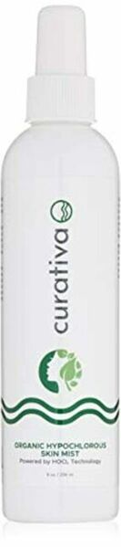
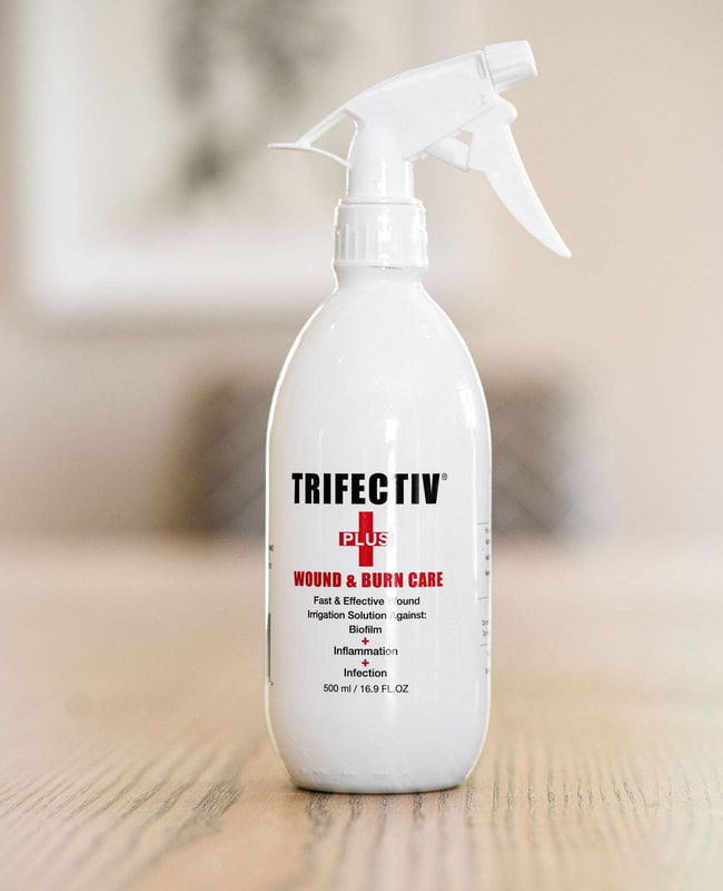
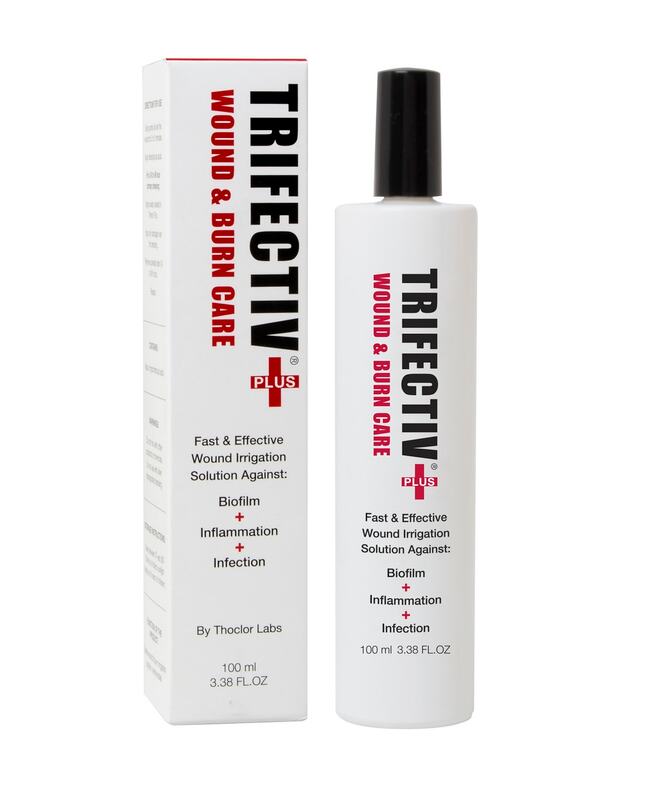
Uses of Hypochlorous Acid
Hypochlorous acid has been investigated as a possible wound care agent,[52][53][54] and as of early 2016 the U.S. Food and Drug Administration has approved products whose main active ingredient is hypochlorous acid for use in treating wounds and various infections in humans and pets. It is also FDA-approved as a preservative for saline solutions.
In a recent study, a saline hygiene solution preserved with pure hypochlorous acid was shown to reduce the bacterial load significantly without altering the diversity of bacterial species on the eyelids. After 20 minutes of treatment, there was >99% reduction of the Staphylococci bacteria.[55]
See also
References
Novel Coronavirus (2019-nCoV) – Aqualution, Effective Prevention and Control
Posted on February 5, 2020
Novel Coronavirus (2019-nCoV)
Scope for Aqualution Hypochlorous Acid Solution for Effective Prevention and Control
Dr M R Lewis
The current outbreak of novel Coronavirus (2019-nCoV) was first reported in Wuhan, China, on 31 December 2019 and has subsequently reached other regions of China and other countries. As of Monday February 3rd 2020, 2019-nCoV has caused 14,557 confirmed cases (14,411 in China, 146 elsewhere) with 305 deaths (304 in China, 1 in the Philippines). Cases have been confirmed in 23 countries across Asia, Europe and North America and the WHO have stated that the risk is “very high” for China and “high” on a global level (WHO website, 3/2/2020). Early outbreak data largely follows an exponential growth pattern (Zhao et al., 2020). Estimates of the R0 for 2019-nCoV vary with values cited typically between 2.24 and 3.58 (WHO, 2020; Zhao et al., 2020). R0 is the average number of secondary cases generated by a primary case. If R0 <1, an epidemic cannot be sustained; if R0 >1, an epidemic is highly likely (Chowell et al., 2004). The R0 values for SARS and MERS were retrospectively calculated to be 3 and 0.7 respectively, and Ebola 1.5-2 (Chowell et al., 2004).
Coronaviruses belong to one of two subfamilies: Coronavirinae and Torovirinae. They are enveloped viruses typically 120-160µ in diameter. They were first isolated in the 1960s from the nasal cavities of patients suffering with colds. Coronaviruses are said to be responsible for 10-15% of common colds worldwide an have a seasonal pattern (the R0 value for seasonal influenza is said to be 1.3). They primarily affect the upper respiratory tract of mammals and birds. They cause direct (viral) and secondary (bacterial) pneumonia and bronchitis. The strains of Coronavirus known to affect humans are shown in Table 1.
Coronaviruses, as with Hepatitis, Influenza, Herpes, Newcastle Disease and Orthopox, are enveloped viruses. Enveloped viruses are viruses that possess an envelope or outer coating that is composed of a lipid layer (fat-like substance that is water insoluble). The envelope is needed to aid in attachment of the virus to the host cell. Loss of the envelope results in loss of infectivity. The mode of transmission for enveloped viruses is characterised by the specific virus; however, the most common routes are via indirect or direct contact of infectious virus particles, contact with or inhalation of respiratory droplets. Some enveloped viruses may also be transmitted by airborne transmission. Enveloped viruses are easily inactivated by routine surface cleaning and disinfection. The inactivation mechanisms of several common virucidal agents have been reported. Treatments with ultraviolet (UV) radiation and Aqualution hypochlorous acid usually destroy the viral genome, whereas chlorine dioxide and heat interrupt the process of host cell recognition for virus binding (Wigginton, 2012).
Table 1: The seven strains of Coronavirus known to affect humans
VariantGenusYear first reportedReported number of
cases / deaths / mortality rate
Human CoV 229EAlphaCoronavirus––
Human CoV OC43BetaCoronavirus––
SARS-CoVBetaCoronavirus20038098 / 774 / 9.6%
Human CoV NL63AlphaCoronavirus2004–
Human CoV HKU1BetaCoronavirus2005–
MERS-CoVBetaCoronavirus20122182 / 779 / 36%
2019-nCoVBetaCoronavirus201914557 / 305 / 2.1% (as of 3/2/2020)
For many years, epidemiologists were only aware of two strains of Coronavirus (Human CoV 229E and Human CoV OC43). Resultant illnesses is fairly mild, affecting just the upper airway. The discovery of SARS-CoV in China added a third. By the end of 2005 two more (mild illness) strains had been identified (Human CoV NL63 and Human CoV HKU1).
Middle Eastern Respiratory Syndrome Coronavirus (MERS-CoV) was identified in 2012 and the new variant (as yet unnamed) 2019-nCoV was first observed in Wuhan, China in late 2019.
Coronaviruses cause a wide range of disease in farm animals and domesticated pets: porcine CoV, bovine CoV (both cause diarrhoea in young animals), avian CoV (respiratory tract), Canine CoV (two types), Feline CoV (two types, both associated with high mortality rates), ferret CoV (two types) and murine CoV (high mortality rate).
Coronaviruses are also present in wild animal species e.g. bats, camels and snakes. Some of these CoV have effective vaccinations (e.g. IBV vaccines are widely used in the commercial poultry industry). These animal diseases are zoonotic and freely mutate, so new viral pandemics continue to pose a very real threat to humans and animals. Consequently, there is much academic and government interest in emerging viruses.
The term “emerging viruses” is used to describe the appearance of viruses whose presence has increased over the past twenty years or whose presence threatens to increase in the years to come (Artika and Ma’roef, 2017).
Emerging viruses include those that have been newly diagnosed in the civil population or those that may have been present before but have mutated. High profile viruses that meet this definition include the highly pathogenic avian influenza (HPAI) virus of subtype H5N1, severe acute respiratory syndrome (SARS-CoV), Ebola, the Middle East respiratory-syndrome Coronavirus (MERS-CoV), Zika and most recently the new variant Wuhan Coronavirus (2019-nCoV). Diseases caused by emerging viruses threaten human and animal health. The majority of emerging viruses are zoonotic. Their appearance is believed to be driven by a number of factors such as socio-economic, environmental and ecological changes (Jones et al., 2008). More local interaction with wildlife in undeveloped countries, greater levels of global travel and trade, and different land use have also been identified as contributing factors for their rapid emergence. Such factors, together with a substantial increase of human population over the past five decades, and urbanisation in developing countries, have contributed to the increased chance of viral diseases emergence and re-emergence.
Of the six previously known strains, two have been associated with serious outbreaks: SARS and MERS.
SARS-CoV was first reported in China in 2002 and caused severe illness (muscle ache, headache, fever followed by respiratory symptoms including pneumonia). WHO state that approximately 25% of cases were seen in health care professionals due to the highly contagious nature of the virus. Whilst most of the cases were in China, it affected over 30 countries. No new outbreaks have been reported since 2014.
MERS-CoV was first discovered in 2012 in Saudi Arabia. Although it has been reported in 27 countries, 85% of the cases have been in Saudi Arabia. Infected patients present with a range of symptoms including fever, shortness of breath, cough, nausea, headache, vomiting and diarrhoea. Epidemiological data suggest that those affected are predominantly males who also suffer with co-morbid disease (eg diabetes) (Hui et al., 2018).
SARS-CoV has been shown to persist on hard surfaces for up to 96 hours (Duan et al., 2003) and up to 5 days if dry (Rabenau et al., 2005b); MERS-CoV persists for at least 48 hours on surfaces (Hui et al., 2018). Environmental contamination is considered a serious reservoir of infection in hotels, public places and health care settings (Radun et al., 2003). Environmental contamination by MERS-CoV in patients’ rooms has been reported in South Korea, with positive MERS-CoV RT-PCR results for cultures from environmental swabs taken from bed sheets, bed rails, intravenous fluid hangers and tables (Bin et al., 2016). In the same study viable MERS-CoV viruses could still be isolated from three of the four enrolled patients studied on days 18–25 after symptom onset. Another study (Cho et al., 2016) detected the presence of MERS-CoV by RT-PCR in viral cultures from four of seven air samples taken from two patients’ rooms, one patient’s bathroom, and one common corridor. MERS-CoV was also detected in the viral cultures for 15 of 68 surface swabs. A third study showed low concentrations of MERS-CoV RNA for environmental swabs taken from bed guardrails and monitors. Even after cleaning the monitors with 70% alcohol based disinfectant, RT-PCR showed low concentrations of MERS-CoV RNA remained: the samples only became negative for MERS-CoV after the monitors were wiped with diluted sodium hypochlorite (Song et al., 2015). This is consistent with the findings of other researchers eg Rabenau et al. (2005) found that (under laboratory conditions) alcohol gels were effective against feline calicivirus in the presence of fetal calf serum but not effective in the presence of sheep erythrocytes or serum albumin: clearly alcohol gels are not efficacious under all “dirty” conditions.
Inanimate surfaces can be as infective as people so prudent and correct use of disinfectants is paramount in environmental control. SARS-CoV can be inactivated (4 log reduction) “quite easily” with many commonly used disinfectants (Rabenau et al., 2005). These workers reported a 30 – 60 minute contact time at in use concentrations of disinfectants based on glutaraldehyde and benzalconium chloride. They did not test any chlorine based products. Dellano et al. (2009) tested a range of household disinfectants against Murine Hepatitis Virus (a SARS-CoV surrogate), including sodium hypochlorite. They found that a 30 second contact time was sufficient to achieve a 4.5 log reduction of MHV dried onto stainless steel coupons. Fogging has also been tested as a viricidal vehicle for disinfectants: Knotzer and workers (2015) fogged stainless steel coupons with dry residue of a range of viruses (enveloped and non-enveloped). They demonstrated that 5 minutes fogging with hydrogen peroxide / peracetic acid (at a rate of 0.4ml/m3) was sufficient to achieve inactivation. However, this disinfectant is very aggressive and, if not used in the correct quantity can be very dangerous to humans. Aqualution hypochlorous acid is safe for humans and the environment. Published work (Clark et al., 2006) has demonstrated excellent log reductions of up to 107 against MRSA (on ceramic tiles). Our informal studies have repeatedly demonstrated that Aqualution products are effective against bacteria and viruses in a wide range of applications (e.g. food safety, animal production, health care) and methods of use (e.g. spray, dip, fog).
One of the most commonly studied viruses is Feline calicivirus (FCV) as it is relatively easy and safe to work with under laboratory conditions. It is also used as a surrogate for norovirus. Both norovirus and FCV are non-enveloped viruses which are harder to kill than enveloped viruses such as Coronaviruses. Many studies have reported on various compounds used for the inactivation of FCV, including acids and alcohols (Whitehead and McCue, 2010), ozone gas (Hudson et al., 2007), H2O2 vapors, and chlorine dioxide gas (ClO2) (Morino et al., 2009) . Whitehead and McCue (2010) showed that bleach and acid-based disinfectants could inactivate FCV within 1 min (>4 log10 reduction). The use of ClO2 has been shown to reduce FCV titers by >3 log10 within 10 h (Morino et al., 2009), and ozone can inactivate FCV in less than 1 h (Hudson et al., 2007). Some of these compounds are toxic, some expensive and most require an extended time for virus inactivation. Aqualution hypochlorous acid (150ppm) achieved >5 log reduction in FCV titres in less than a minute (Chander et al., 2012).
Various virucides are commonly used to disinfect environmental contact surfaces implicated in viral outbreaks. The material safety data sheets and labels for these virucidal compounds rarely allow for their aerosolization, spraying, or fogging due to their toxicity and adverse health effects for given exposure durations and concentrations. Many of these chemical compounds, such as sodium hypochlorite, chlorine gas, and glutaraldehyde, have been associated with occupational illnesses. For example, exposure to glutaraldehyde is associated with contact dermatitis in health workers, and the use of quaternary ammonium compounds has been found to cause occupational asthma in users (Purohit et al., 2000; Ravis et al., 2003). For cases in which aerosolization is approved, the use of personal protective equipment and a self-contained breathing apparatus is required, which makes the use of these compounds difficult, especially in public places such as hospitals or schools. Aqualution hypochlorous acid has been demonstrated as a powerful disinfectant and has been shown to be efficacious against a wide range of microorganisms in solution and when sprayed in the air. Another significant benefit of Aqualution hypochlorous acid is its lack of toxicity at ready-to-use (RTU) concentrations. In-house testing has repeatedly shown that fogging of Aqualution RTU Aqualution hypochlorous acid products achieves safe and consistent log reductions in bacterial and viral loading, both on environmental surfaces (eg aged care facilities) and food products (eg eggs, fresh raspberries).
SARS-CoV and MERS-CoV were emerging diseases so no medical treatment or vaccines were available and control of the epidemics relied on rapid diagnosis, isolation of patients and attention to infection control. This will also be the case for any future pandemics caused by emerging viruses. According to WHO (2015) cleaning surfaces with water and detergent followed by application of a commonly used disinfectant is an effective and sufficient procedure to ensure good environment hygiene.
Rutala and Weber (2014) proposed a hierarchy approach to anticipating the effectiveness based on the general resistance to disinfection of various microorganisms, shown in the table below.
Table 2: Hierarchy of Microbial Resistance to Disinfectants and Sterlilants
Most ResistantMicroorganismAqualution BS EN tests
Spores (eg C. difficile)BS EN 13704:2002; BS EN 13727:2003
Mycobacteria (eg M. tuberculosis)BS EN 14204:2012; BS EN 14348:2005
Non-Enveloped Viruses (eg norovirus, polio)BS EN 14476:2013
Fungi (eg Candida albicans)BS EN 13697:2001; BS EN 1275:1997
Bacteria (eg MRSA, VRE, Acinetobacter)BS EN 1040:2005; BS EN 1276:2009; BS EN 13697:2001; BS EN 1656:2009
Enveloped Viruses (eg HIV, influenza, )BS EN 14476:2013
Most SusceptibleAdapted from Spaulding, 1957 and Rutala and Weber, 2014
In the table above, enveloped viruses are at the bottom of the table in the most susceptible group ie the easiest class of microorganisms to kill. 2019-nCoV is an enveloped virus and falls into this group, therefore we would expect disinfectants effective against this group to also be effective against Coronavirus, when used as per directions of use. We are not currently able to test Aqualution products against 2019-nCoV as the strain is not available for laboratory testing. In addition, even if samples were available, it is not possible to make any definitive claims until regulators review and approve those claims, this is the situation with the newly identified Coronavirus.
However, Aqualution products have passed British Standards tests for virus efficacy and we have conducted our own in-house testing against a wide range of viruses, both enveloped and non-enveloped. We constantly strive to ensure that Aqualution products are efficacious and as such have performed many tests against a wide range of organisms. We have passed all the BS EN tests highlighted in the table above and have not failed to achieve a positive test result to date. We have also carried out tests on a wide range of other organisms to satisfy the very vigorous demands of the Biocidal Products Regulation (BPR) under which our active substance is approved and for which we own the dossiers submitted to ECHA regulating Aqualution hypochlorous acid throughout the European Union.
Selecting the most suitable disinfectant is one of the two components essential for effective environmental disinfection. The other component is ensuring that the disinfectant contacts all surfaces, is given the correct dwell time and that manufacturers label instructions are followed. This relies on proper training of hospital staff, especially environmental staff and infection control nurses. The combination of product and practice results in effective surface disinfection, the reduction of patient risk and improved patient outcomes. The five key considerations when selecting a disinfectant are summarized in the table below. The authors of the guidance framework (Rutala and Weber, 2014) suggest giving potential disinfectant products a score in each of the five categories and recommend that the optimal product is the one with the highest score. Aqualution hypochlorous acid manufactured by Aqualution Systems achieves very high scores and as such could offer a safe and efficacious disinfection option in domestic, community and healthcare settings.
Our findings demonstrate that it consistently performs well under both laboratory and field conditions and as such is fit for purpose. We have passed many efficacy tests, including the stipulated BS EN tests. We have achieved the required >3 log reductions in viral loads both under laboratory and commercial “real life” conditions. Alcohol-based sanitizers and disinfectants are effective against viruses in laboratory-based tests, and therefore recommended to prevent viral transmission. However they do not always appear to be effective in “real life” situations. Similarly, the glutaraldehydes tested by Rabenau and workers (2005) achieved good log reductions of viral loading on surfaces. However, their very long contact times (30 -60 minutes) mean that they may be of limited “real world” use as it is not practical nor in some cases safe, to leave them on the surface for that length of time. Aqualution hypochlorous acid achieves >5 log viral reductions in less than a minute and is therefore effective in a spray and wipe cleaning regime.
Aqualution’s Aqualution hypochlorous acid is safe for people and the environment. It can be misted into the environment when people and animals are in place with no deleterious effects. It can be sprayed onto a surface and left with no fear of toxic residues. Workers do not need any PPE to handle it. It can be used on soft and hard surfaces. It is extremely versatile and can be used as a dip, spray, mist or fog. It is simple to use therefore compliance is high. It does not lose efficacy at low temperatures. It has a 12 month shelf life if kept at suitable storage conditions.
CONCLUSION
SARS-CoV, MERS-CoV and 2019-nCoV were and are emerging diseases with no immediate availability of medical treatment or vaccines. This will also be the case for any future pandemics caused by emerging viruses. There is a strong likelihood that other emergent zoonotic viral pandemics will occur in the future. Little evidence is available in the literature concerning inactivation of viruses by disinfectants. What is available indicates that viruses, especially enveloped ones, are relatively easy to inactivate and a wide range of disinfectants may be effective. Important potential areas of contamination / transfer include people (isolation, face masks), animals (hand hygiene), food (thorough cooking, pasteurisation) and the environment (hard surfaces). Rapid case identification, isolation of patients and infection control measures are essential to prevent the spread of these emerging viral diseases within households, the community and health care facilities. Aqualution hypochlorous acid is not specifically tested against 2019-nCoV – no disinfectant is – but it is tested and efficacious against a wide range of microorganisms including spores, bacteria and other viruses (enveloped and non-enveloped) leading to the reasonable conclusion that it will be effective.
Controlling environmental hygiene in homes, public places and health care facilities is paramount. Viruses are able to persist in the environment for days but are relatively easy to kill, especially enveloped ones. Disinfectants need to have complete virucidal activity against both enveloped and non-enveloped viruses. Data on the efficacy of disinfectants against coronovirus are very limited. The BS for virucidal activity (BS EN 14476:2013) uses Adenovirus and Poliovirus as the test organisms, both of which are non-enveloped. Aqualution hypochlorous acid has been tested against viruses and has been shown to have virucidal properties (Pineau, 2000). It has achieved >5 log reductions against orthopoxvirus (enveloped), adenovirus (non-enveloped) and poliovirus (non-enveloped). Disinfectants work best in clean conditions so it is important to clean (with a detergent) then disinfect for optimal results. Although some products pass lab based virucidal tests, they do not work well in “real life” conditions, especially in the presence of soil / organic material. Many products are effective in lab tests but need extended contact times (30 minutes to several hours) to be effective. Again, this is not compatible with “real life” needs for a disinfectant.
Aqualution hypochlorous acid is fast acting with contact times typically less than a minute. It is safe for people and safe for the environment. It is versatile and can be used as a dip, skin irrigant, spray, mist or fog. There may be opportunity to fog areas with disinfectant in an attempt to reduce / inactivate viral load in the air and environment. It is extremely effective against a wide range of micro-organisms, including enveloped viruses. As such it is suitable for use as a broad spectrum, easy to use, efficacious and safe disinfectant.
REFERENCES
Artika, I.M. and Ma’roef, C.N. 2017. Laboratory biosafety for handling emerging viruses. Asian Pacific Journal of Tropical Biomedicine, 7 (5): 483-491.
Bin, S.Y., Heo, J.Y. and Song, M.S. 2016. Environmental contamination and viral shedding in MERS patients during MERS-CoV outbreak in South Korea. Clin Infect Dis. 62: 755–60.
Chander, Y., Johnson, T., Goyal, S.M. and Russell, R.J. 2012. Antiviral activity of Ecasol against feline calicivirus, a surrogate of human norovirus. Journal of Infection and Public Health. 5 (6): 420-424.
Cho, S.Y., Kang, J.-M. and Ha, Y.E. 2016. MERS-CoV outbreak following a single patient exposure in an emergency room in South Korea: an epidemiological outbreak study. Lancet. 388: 994–1001.
Chowell, G., Castillo-Chavez, C., Fenimore, P.W., Kribs-Zaleta, C.M., Arriola, L. and Hyman, J.M. 2004. Model Parameters and Outbreak Control for SARS. Emerging Infectious Diseases. 10 (7): 1258-1263.
Clark, J., Barrett, S. P., Rogers, M. and Stapleton R. 2006. Efficacy of super-oxidized water fogging in environmental decontamination. Journal of Hospital Infection, 64: 386–390.
Dellanno, C., Quinn Vega, M.S. and Boesenberg, D. 2009. The antiviral action of common household disinfectants and antiseptics against murine hepatitis virus, a potential surrogate for SARS Coronavirus. American Journal of Infection Control. 37: 649-52.
Duan, S.M., Zhao, X.S. and Wen, R.F. 2003. Stability of SARS Coronavirus in human specimens and environment and its sensitivity to heating and UV irradiation. Biomedical and Environmental Science, 16:246—255.
Hudson, J.B., Sharma, M. and Petric, M. 2007. Inactivation of norovirus by ozone gas in conditions relevant to healthcare. Journal of Hospital Infection, 66: 40-45
Hui, D.S., Azhar, E.I., Kim, Y.-J., Memish, Z.A., Oh, M.D. and Zumla, A. 2018. Middle East respiratory syndrome Coronavirus: risk factors and determinants of primary, household, and nosocomial transmission. Lancet Infectious Diseases. 18: e217–27.
Jones, K.E., Patel, N.G., Levy, M.A., Storeygard, A., Balk, D., and Gittleman, J.L. 2008. Global trends in emerging infectious diseases. Nature. 451: 990-993.
Knotzer, S, Kindermann, J, Modrof, J and Kreil, T.R. 2015. Measuring the effectiveness of gaseous virus disinfectants. Biologicals 43, 519-523
Morino, H., Fukuda, T., Miura, T., Lee, C., Shibata, T. and Sanekata, T. 2009. Inactivation of feline calicivirus, a norovirus surrogate, by chlorine dioxide gas. Biocontrol Science, 14: 147-153
Purohit, A., Kopferschmitt-Kubler, M.C., Moreau, C., Popin, E., Blaumeiser, M. and Pauli, G. 2000. Quaternary ammonium compounds and occupational asthma.
International Archives of Occupational and Environmental Health, 73: 423-427.
Rabenau, H.F, Kampfb, G., Cinatla, J and Doerr, H.W. 2005. Efficacy of various disinfectants against SARS Coronavirus. Journal of Hospital Infection, 61, 107–111
Rabenau HF, Cinatl J, Morgenstern B, Bauer G, Preiser W, and Doerr, H.W. 2005b. Stability and inactivation of SARS Coronavirus. Medical Microbiology and Immunology. 194, 1—6.
Radun D, Niedrig M, Ammon A, and Stark K. 2003. SARS: retrospective cohort study among German guests of the hotel “M”, Hong Kong. Eurosurveillance. 8:228—230.
Ravis, S.M., Shaffer, M.P., Shaffer, C.L., Dehkhaghani, S. and Belsito, D.V. 2003. Glutaraldehyde-induced and formaldehyde-induced allergic contact dermatitis among dental hygienists and assistants. Journal of the American Dental Association, 134: 1072-1078
Rutala, W.A. and Weber, D.J. 2014. Selection of the ideal disinfectant. Infection Control and Hospital Epidemiology. 35 (7): 855-865.
Song, J.Y., Cheong, H.J. and Choi, M.J. 2015. Viral shedding and environmental cleaning in Middle East respiratory syndrome Coronavirus infection. Infect Chemotherapy. 47: 252–55.
Spaulding, E.H. 1957. Chemical disinfection and antisepsis in the hospital. J Hosp Res, 9: 5-31.
Whitehead, K. and McCue, K.A. 2010. Virucidal efficacy of disinfectants active against feline calicivirus, a surrogate for norovirus, in a short contact time
American Journal of Infection Control, 38: 26-30
WHO. 2009. WHO guidelines on natural ventilation for infection control in health-care settings. Geneva: World Health Organization, 2009.
WHO. 2015. Infection prevention and control during health care for probable or confirmed cases of Middle East respiratory syndrome Coronavirus (MERS-CoV) infection Interim guidance. Geneva: World Health Organization, 2015.
Wigginton, K.R., Pecson, B.M., Sigstam, T., Bosshard, F. and Kohn, T. 2012. Virus inactivation mechanisms: impact of disinfectants on virus function and structural integrity. Environmental Science and Technology, 46: 12069-12078.
Zhao, S., Lin, Q., Ran, J., Musa, S.S., Yang, G., Wang, W., Lou, Y., Gao, D., Yang, L., He, D. and Wang, H.W. 2020. Preliminary estimation of the basic reproduction number of novel coronavirus (2019-nCoV) in China, from 2019 to 2020: A data-driven analysis in the early phase of the outbreak. International Journal of Infectious Diseases, [In press].
